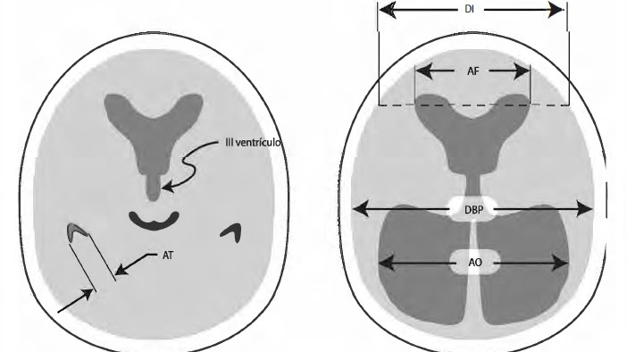
DI = Internal diameter, AF= Frontal horns, DBP = Biparietal diameter,
AO = Occipital horns, AT = Temporal horns
EVANS INDEX
A result > 0.3 = Hydrocephalus
AF/DI QUOTIENT
< 40%: normal
40% to 50%: borderline
> 50%: suggestive of hydrocephalus.
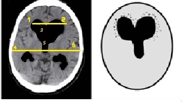
1-2 Ballooning of the frontal horns
5 Ballooning of the third ventricle
“Mickey Ears Sign”“
OTHER IMAGING CRITERIA FOR HYDROCEPHALUS
The size of the temporal horns is 2 mm wide; if there is no hydrocephalus, the temporal horns are barely visible.
Ballooning of the frontal horns and lateral ventricles (Mickey Mouse figure)
Ballooning of the third ventricle, under normal conditions it has a similar appearance to a slit ventricle.
Periventricular hypodensity on CT or periventricular hyperintensity on T2-weighted MR images (transependymal edema)